Ct Anatomy Pelvis Muscles / Radiologia Brasileira Procedimentos Percutaneos Pelvicos Guiados Por Imagem Revisao Das Principais Vias De Acesso - This mri male pelvis axial cross sectional anatomy tool is absolutely free to use.
Ct Anatomy Pelvis Muscles / Radiologia Brasileira Procedimentos Percutaneos Pelvicos Guiados Por Imagem Revisao Das Principais Vias De Acesso - This mri male pelvis axial cross sectional anatomy tool is absolutely free to use.. Females' pelvis is wider and the pubis shorter than males'. Innervation of the female levator ani muscles. Axial mr high resolution (small fov). Attached to the pelvis are muscles of the buttocks, the lower back, and the thighs. Anatomy of the thorax, heart, abdomen and pelvis the following video will go through normal abdominal anatomy on ct imaging.
Learn about anatomy muscles pelvis with free interactive flashcards. Anatomy of the thorax, heart, abdomen and pelvis the following video will go through normal abdominal anatomy on ct imaging. Females' pelvis is wider and the pubis shorter than males'. The muscles are connected with the bones. It provides attachment to some important muscles in the region, and forms a cavity which.
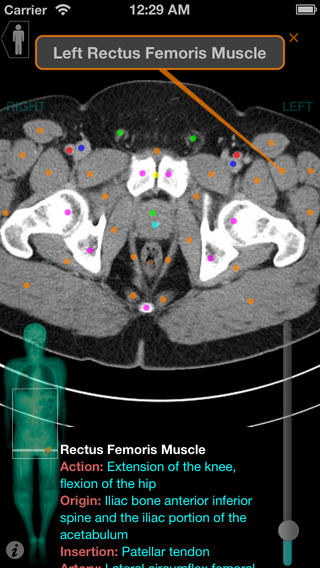
X Anatomy Mobile App Editors Pick from 0f14676b303fd91881eb-98dd17e178263eba3c55ca6434a72b9d.ssl.cf5.rackcdn.com Muscles of the pelvis that cross the lumbosacral joint to attach onto the trunk were described in the previous blog post note: Functional anatomy of the male pelvicfloor explore the important aspects of the structures and functions of the male pelvic. If you want to learn how to read ct scans of the abdomen and pelvis proficiently, this video is an excellent starting point. Anatomy pelvis muscles pubococcygeus, puborectalis and iliococcygeus., pelvis nerve, the spinal nerves that arise from vertebral column through the sacrum., pelvic floor musculature laminated anatomy anatomy pelvis muscles; This is the sixth in a series of 8 blog post articles on the anatomy and physiology of the lumbar spine and pelvis. The muscles are connected with the bones. Abdominal and pelvic anatomy encompasses the anatomy of all structures of the abdominal and pelvic cavities. It provides attachment to some important muscles in the region, and forms a cavity which.
Abdominal and pelvic anatomy encompasses the anatomy of all structures of the abdominal and pelvic cavities.
Pelvic floor muscles that are located wholly within the pelvis. Architectural differences in the bony pelvis of women with and without pelvic floor disorders. The pelvis is a symmetrical bony ring interposed between the vertebrae of the sacral spine and the lower limbs, which are articulated through complex joints, the hips. This is the iliopubic line which outlines the anatomic anterior column this is the ilioischial line which outlines the anatomic posterior column. N patient preparation n patient position n scanogram. The lateral superficial muscles, the transversus and external and internal oblique muscles, originate on the rib cage and on the pelvis (iliac crest and inguinal ligament) and are attached to the anterior and posterior layers of the sheath of the rectus. We study anatomy at the practical anatomy class we study the human body. It attaches to the walls of the lesser pelvis, separating the pelvic cavity from the perineum inferiorly (region which includes the in this article, we shall look at the anatomy of the muscles that make up the inferior lining of the cavity; It affects the entire lower limb and the movement of the hip and the lumbar area. Ischial tuberosity which flexor of the knee attaches here? Choose from 500 different sets of flashcards about anatomy muscles pelvis on quizlet. Axial pelvis ct axial femur ct axial femur ct axial knee ct. Use the mouse scroll wheel to move the images up and down alternatively use the tiny arrows (>>) on both side of the image to move the images.
Females' pelvis is wider and the pubis shorter than males'. The gastrocnemius muscle is a complex muscle that is fundamental for walking and posture. Abdominal and pelvic anatomy encompasses the anatomy of all structures of the abdominal and pelvic cavities. The pelvis is a basin shaped bony structure formed by the combination of two pelvic bones (hip bones or innominate bones) and the sacrum. Axial mr high resolution (small fov).
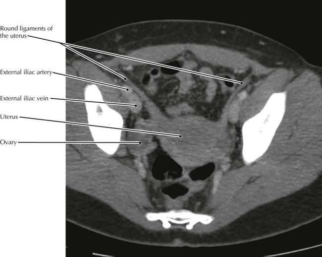
Pelvis And Perineum Radiology Key from radiologykey.com Anatomy pelvis muscles pubococcygeus, puborectalis and iliococcygeus., pelvis nerve, the spinal nerves that arise from vertebral column through the sacrum., pelvic floor musculature laminated anatomy anatomy pelvis muscles; Hepatocellular carcinoma or liver cancer. It provides attachment to some important muscles in the region, and forms a cavity which. There are many muscles that form the pelvic floor, including puborectalis, pubococcygeus, iliococcygeus and coccygeus. Innervation of the female levator ani muscles. The full bladder displaces small bowel loops superiorly. Ct anatomy of the pelvis. Anatomical drawing of the female pelvis.
This anatomy section promotes the use of the terminologia anatomica, the international standard of anatomical nomenclature.
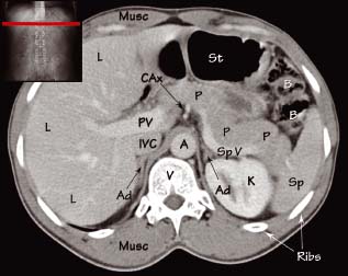
It attaches to the walls of the lesser pelvis, separating the pelvic cavity from the perineum inferiorly (region which includes the in this article, we shall look at the anatomy of the muscles that make up the inferior lining of the cavity; Anatomy of the thorax, heart, abdomen and pelvis the following video will go through normal abdominal anatomy on ct imaging. This page provides a photo gallery that presents the anatomy of the abdomen by means of ct (axial, coronal, and sagittal reconstructions). • to assess equivocal imaging findings • staging of hepatic neoplasms • metastatic workup of primary malignancies • diagnosis of abdominal masses • assessment of biliary problems • diagnosis of vascular lesions. Muscles of the pelvis that cross the lumbosacral joint to attach onto the trunk were described in the previous blog post note: The lateral superficial muscles, the transversus and external and internal oblique muscles, originate on the rib cage and on the pelvis (iliac crest and inguinal ligament) and are attached to the anterior and posterior layers of the sheath of the rectus. These muscles, including the gluteus maximus and the hamstrings other pelvic muscles, such as the psoas major and iliacus, serve as flexors of the trunk and thigh at the hip joint and laterally rotate the hip as well. Hepatocellular carcinoma or liver cancer. Females' pelvis is wider and the pubis shorter than males'. Architectural differences in the bony pelvis of women with and without pelvic floor disorders. Labeled scrollable mri of the pelvis covering anatomy with a level of detail appropriate for medical students. Axial section through male bladder. If you want to learn how to read ct scans of the abdomen and pelvis proficiently, this video is an excellent starting point.
We'll go through the on this image, we can also see some of the muscles that we talked about specifically the slowest. Anatomical drawing of the female pelvis. Renal pelvis or ureter cancer. Axial mr high resolution (small fov). Labeled scrollable mri of the pelvis covering anatomy with a level of detail appropriate for medical students.
Abdominal Ct Anatomy Radiology Key from radiologykey.com The muscles are connected with the bones. Learn about anatomy muscles pelvis with free interactive flashcards. They support the pelvic organs especially during increases in intra abdominal pressure and also aid in urinary and faecal. We'll go through the on this image, we can also see some of the muscles that we talked about specifically the slowest. Hepatocellular carcinoma or liver cancer. Males and females differ significantly in the anatomy of the pelvis: This mri male pelvis axial cross sectional anatomy tool is absolutely free to use. Muscles of the pelvis that cross the lumbosacral joint to attach onto the trunk were described in the previous blog post note:
Ischial tuberosity which flexor of the knee attaches here?
They support the pelvic organs especially during increases in intra abdominal pressure and also aid in urinary and faecal. 13 what portion of the bony pelvis is the arrow pointing to? Rib thorax lumbar pelvis sacrum coccyx femur fibula tibia. It affects the entire lower limb and the movement of the hip and the lumbar area. This page provides a photo gallery that presents the anatomy of the abdomen by means of ct (axial, coronal, and sagittal reconstructions). This mri male pelvis axial cross sectional anatomy tool is absolutely free to use. Figure 6.4 • ct scan of pelvis: Females' pelvis is wider and the pubis shorter than males'. Hepatocellular carcinoma or liver cancer. Abdominal and pelvic anatomy encompasses the anatomy of all structures of the abdominal and pelvic cavities. It is strengthened and supported by several joints and ligaments. The lateral superficial muscles, the transversus and external and internal oblique muscles, originate on the rib cage and on the pelvis (iliac crest and inguinal ligament) and are attached to the anterior and posterior layers of the sheath of the rectus. Axial section through male bladder.
Anatomy of the thorax, heart, abdomen and pelvis the following video will go through normal abdominal anatomy on ct imaging anatomy muscles pelvis. N patient preparation n patient position n scanogram.


0 Komentar